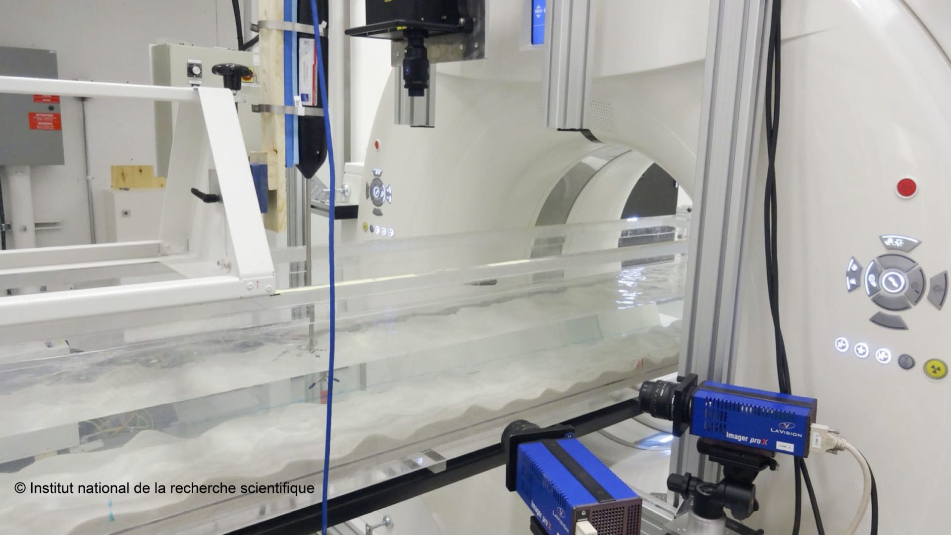
Photo courtesy of Institut national de la recherche scientifique (INRS) – On the photo: the two blue cameras are high-speed cameras that track the movement of particles in water, the channel being illuminated by a laser (the black box seen above). In scientific jargon, this device is known as a PIV (particle image velocimetry) system. The last device plunged into the water on the far left is a Vectrino, a Doppler current meter. This image was taken as part of the experiments whose results were shared in this publication: https://ascelibrary.org/doi/10.1061/%28ASCE%29HY.1943-7900.0002025
A medical imaging technique known as the computed tomography or CT scan is now being used to scan sections of tree trunks, sediment cores, minerals and archaeological objects, so that their interiors can be examined without having to destroy them.
Philippe Després, a researcher in the Department of Physics, Physical Engineering and Optics at the Université Laval and at the Institut universitaire de pneumologie et de cardiologie de Québec, worked with the team led by Pierre Francus, a professor at the Institut national de la recherche scientifique, to adapt and accelerate the mathematical operations that drive computed tomography. These algorithms are used to reconstruct 3D images from a series of one-dimensional and two-dimensional X-ray projections.
CT scanning was originally designed to obtain internal images of the various parts of the human body which, with the exception of bone, is primarily composed of low-density organs and tissues. When this technique is used to scan a sediment core (layers of particles such as sand) or high-density mineral samples, the image obtained often contains artifacts—errors—that limit analysis.
Philippe Després and his colleagues have therefore developed image reconstruction algorithms adapted to high-density objects, making it possible to obtain images that better represent the interior of such samples.
These new algorithms facilitate the interpretation of images obtained in the laboratory using a hydraulic channel, which provides a better understanding of sediment-current interactions. Better still, they could eventually be used in medicine to improve image quality in patients with a prosthetic hip or other metal implant, as these materials produce artifacts that interfere with the interpretation of medical results.
Video provided by Mr. Pierre Francus, Professor at the Institut national de la recherche scientifique (INRS)